
About Uterine Fibroid Embolization
Uterine fibroid embolization, or UFE , is performed by interventional radiologists, who consult with a woman’s gynecologist to evaluate whether UFE is right for the patient. Interventional radiologists are physicians who use X-rays and other imaging techniques to see inside the body and guide tiny catheters and other micro-tools to perform fibroid embolization and treat other conditions without surgery.
Uterine fibroid embolization is not performed by gynecologists, who, for various reasons, may sometimes not discuss it as a possible option for their patients with fibroid tumors. This may occur because gynecologists are not familiar with the procedure or the favorable results of clinical studies. Sometimes they are unaware that UFE is an FDA-approved treatment for fibroid tumors, and may mistakenly tell patients that it is “experimental”.
More information is available on the ACOG site.
In the past, there also were problems with insurance companies, especially HMOs who would not pay for a “new” procedure. This is rarely a problem today and most insurers recognize uterine fibroid embolization as an effective, approved procedure that often is safer and less costly than other options.
Advantages of Uterine Fibroid Embolization
UFE has several advantages over the other treatments for uterine fibroids.
- It involves no blood loss, no stitches or surgical incision (only a small nick in the skin), no abdominal scar, no general anesthesia, no prolonged hospital stay, and no burning or scraping the lining of the uterus.
- Uterine fibroid embolization requires much less time off work than hysterectomy (a few days to a week, versus six weeks or more).
- One advantage compared to myomectomy is that uterine fibroid embolization can be used to treat all fibroids at the same time, regardless of their size or location in the uterus.
- Unlike treatment with Lupron, premature symptoms of menopause are not usually induced. The materials used for uterine fibroid embolization are well tolerated and FDA-approved for embolization.
- The cost of UFE varies, but is comparable to or less than the cost of hysterectomy or myomectomy.
Papers on this topic are available at these professional publication sites:
Hysterectomy paper | Myomectomy paper (links to publication sites)
In the few cases that fibroid embolization is not successful and surgery is needed, the operation is typically easier after UFE, with much less bleeding.
Because UFE is new, long-term data on the recurrence rates of treated fibroids are not yet available.
Although uterine fibroid embolization is a relatively new treatment for fibroids, the same procedure has been used with great success to treat emergency uterine bleeding for more than 25 years. When the procedure is used to control bleeding, it is generally referred to as Uterine Artery Embolization.
See medical references on Uterine Fibroid Embolization.
How Uterine Fibroid Embolization is Performed
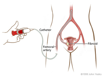
Uterine fibroid embolization blocks, or “embolizes” the blood vessels that “feed” the uterine fibroid, causing it to shrink. The interventional radiologist performs the fibroid embolization through a tiny tube called a catheter which is inserted into an artery at the top of the leg (Figure 1). The patient is given a local anesthetic to numb the skin and a mild sedative, so the procedure is not painful. A special X-ray machine that creates moving pictures in “real” time enables the doctor to see the catheter as it is guided through the blood vessels and into the uterine artery. A contrast agent, or dye, is injected to highlight the blood vessels in the uterus to create an “arteriogram,” an X-ray that maps the arteries feeding the fibroids.
Figure1
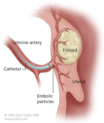
The final step in fibroid embolization is the injection of tiny particles through the catheter. The particles lodge in the blood vessels feeding the fibroids and cut off their blood supply (Figure 2), but the uterus and ovaries are spared. After uterine fibroid embolization, the fibroids begin to shrink. The catheter is removed and the patient is observed overnight, and usually goes home the next day. The fibroids continue to shrink for several months after fibroid embolization.
Figure 2
Click for an animation of the uterine fibroid embolization procedure.